○Key Points:
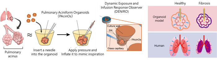
◆We developed Pulmonary Aciniform Organoids (PAcinOs), derived from human iPS cells, that mimic the peripheral lung structure known as the acinus. We also created a custom device, the Dynamic Exposure and Infusion Response Observer (DENIRO), which enables direct intraluminal access to organoids.
◆By combining these technologies (PAcinOs in DENIRO), we were able to inflate and observe the biomechanics of this lung-like microstructure that mimics breathing. This allowed us, for the first time, to quantitatively measure lung compliance -- the stiffness of lung organoid.
◆This system may enable modeling of lung-stiffening diseases, such as pulmonary fibrosis, and facilitate evaluation of candidate therapeutic drugs.
A new technique to inflate lung organoids from within
○Overview:
A research group led by Shoji Takeuchi, Project Professor at the Institute of Industrial Science, The University of Tokyo (Professor at the Graduate School of Information Science and Technology, The University of Tokyo), Satoshi Ikeo, Project Assistant Professor at the Institute of Industrial Science, The University of Tokyo, Yuta Tani, a graduate student at the Graduate School of Information Science and Technology (at the time of the study), and Yuki Yamamoto, CEO of HiLung Inc., developed pulmonary aciniform organoid (PAcinO) (Note 1), which mimics the peripheral lung structure known as the pulmonary acinus. The team also developed a device called DENIRO that enables direct access to the interior of organoids and allows pressure to be applied from the lumen.
By integrating these technologies, the researchers established an experimental platform called PAcinOs in DENIRO. Using non-invasive three-dimensional imaging with optical coherence tomography (OCT) (Note 2), they successfully quantified organoid volume changes in response to pressure and measured lung compliance (Note 3). In addition, by treating PAcinOs with bleomycin (Note 4), a drug known to induce fibrosis, they reproduced fibrosis-like mechanical stiffening in the organoids and evaluated the effects of an existing drug used to treat pulmonary fibrosis.
Conventional two-dimensional culture systems cannot adequately reproduce three-dimensional structures or mechanical functions. Meanwhile, typical organoid cultures are either unable to replicate an inner lumen, or even if they do, still are not accessible due to its size limitations. In this study, the researchers utilized human iPS cell-based organoid engineering and constructed grape-like structures that are sufficiently large and resemble the architecture of the actual lung, making intraluminal access possible. By precisely controlling pressure through this access, the platform enables evaluation of dynamic mechanical responses while preserving the three-dimensional structure.
These findings provide platform technology that enables dynamic evaluation of structure and biomechanics of lungs which are extremely difficult to observe in a living body. The system is expected to contribute to a better understanding of diseases characterized by lung stiffening, such as pulmonary fibrosis, and to facilitate evaluation of potential therapeutic drugs.
○Comment from the Researcher Shoji Takeuchi: "A possible future": The lung is an organ that naturally expands and contracts with each breath. While conventional organoids have been able to reproduce lung morphology, they have not been able to reproduce its movement. We felt that a lung model that does not move is somehow incomplete, so we attempted to introduce motion by applying pressure from inside the organoid. I still remember the moment when the organoids expanded beautifully, the entire lab cheered. By giving the organoids this dynamic behavior, we believe it will become possible to study lung function and disease states in conditions that more closely resemble those in the human body. In the future, beyond applications in medicine and drug discovery, we hope this technology could also contribute to new systems such as respiratory components for biohybrid robots.
○Research Details:
The lung is an essential organ responsible for gas exchange, taking in oxygen and releasing carbon dioxide. In the peripheral region of the lung are numerous grape-like microscopic structures called pulmonary acini. With each breath, the lung repeatedly expands and contracts, deforming flexibly to enable efficient gas exchange. This flexibility is evaluated by a mechanical parameter known as lung compliance. In pulmonary fibrosis, severe respiratory diseases, lung tissue becomes stiff and lung compliance decreases.
Previous studies have developed lung-on-a-chip models (Note 5) to mimic such disease conditions in vitro. However, they cannot fully reproduce the three-dimensional structure of the lung because these systems rely mainly on two-dimensional culture. On the other hand, conventional spheroid organoid culture systems can replicate alveolar curvature and luminal cellular characteristics found in vivo, but they remain static systems that cannot accommodate mechanical stimulation or provide access to the internal lumen. Therefore, there has been a strong need for a model that preserves the unique three-dimensional structure of the lung while also reproducing its dynamic behavior in vitro.
In this study, inspired by the peripheral structure of the human lung, the research team constructed larger Pulmonary Aciniform Organoids (PAcinOs) by fusing multiple spheroids (Note 6). In addition, the researchers developed a custom device made of polydimethylsiloxane (PDMS) (Note 7), called the Dynamic Exposure and Infusion Response Observer (DENIRO), which facilitates access to the interior of organoids. The team successfully cultured PAcinOs on this device (Fig. 1).
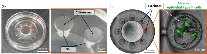
Fig. 1 PAcinOs in DENIRO system
(A) The custom device DENIRO, equipped with eight culture wells containing slits that allow intraluminal access.
(B) PAcinOs cultured on DENIRO. The organoids contain alveolar epithelial type II cells, an important tissue stem cell population in the lung.
Using this PAcinOs in DENIRO system, the researchers established a method to gradually apply pressure from the lumen of the organoid. While performing non-invasive three-dimensional imaging using optical coherence tomography (OCT), an extremely fine glass capillary was inserted through a slit in the culture well to puncture the PAcinO and deliver controlled pressure from within (Fig. 2A, B). The procedure was performed in three groups: untreated PAcinOs (normal model), bleomycin-treated PAcinOs (fibrosis model), and PAcinOs treated with both bleomycin and the clinically-approved pulmonary fibrosis drug nintedanib (treatment model). Pressure loading was successfully applied to organoids in all three conditions (Fig. 2C).

Fig. 2 Intraluminal access and pressure application using the PAcinOs in DENIRO system
(A) Schematic illustration of intraluminal access using the PAcinOs in DENIRO system.
(B) Insertion of a glass capillary into the lumen of PAcinOs while being observed with OCT.
(C) Organoids expanding in response to pressure loading in PAcinOs under each condition.
By continuously measuring the volume changes of PAcinOs during pressure loading and dividing the change in volume by the applied pressure difference, the researchers succeeded, for the first time, in quantifying lung compliance in organoid lungs (Fig. 3). In fibrosis-model PAcinOs, the organoids did not expand even when pressure was applied, and lung compliance was significantly lower than that in normal-model PAcinOs. In treatment-model PAcinOs, lung compliance tended to recover, although the difference was not statistically significant. These results suggest that dynamic parameters such as lung compliance can be used in vitro to evaluate disease states and drug effects using organoid models.
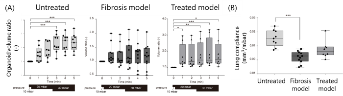
Fig. 3 Pressure-induced volume change and lung compliance of PAcinOs under each condition
(A) Volume changes of organoids as pressure was increased every 2 minutes 30 seconds. Untreated normal PAcinOs expanded at approximately 20-30 mbar, whereas expansion was not clearly observed in fibrosis-model or treatment-model PAcinOs.
(B) Lung compliance in each group. Fibrosis-model PAcinOs showed reduced lung compliance compared with normal PAcinOs.
These results demonstrate platform technology that enables simultaneous evaluation of structure and mechanical properties in a human iPSC-derived three-dimensional lung model. This approach is expected to contribute to a better understanding of diseases characterized by lung stiffening, such as pulmonary fibrosis, and to facilitate the evaluation of potential therapeutic drugs.
○Researchers:
Shoji Takeuchi
Project Professor, Institute of Industrial Science, the University of Tokyo
Professor, Grad. School of Information Science and Technology, the University of Tokyo
Satoshi Ikeo
Project Research Associate, Institute of Industrial Science, the University of Tokyo
Yuta Tani
Master's student, Grad. School of Information Science and Technology, the University of Tokyo (at the time of the study)
Yuki Yamamoto
CEO, HiLung Inc.
○Publication Information:
Journal: Biomaterials
Title: Dynamic in vitro platform for mechanical profiling of human pulmonary aciniform organoids via intraluminal access
Authors: Satoshi Ikeo, Yuta Tani, Jun Sawayama, Shogo Nagata, Harry Choi, Toshio Suzuki, Saburo Ito, Tetsuharu Nagamoto, Yuki Yamamoto*, Shoji Takeuchi*
DOI: 10.1016/j.biomaterials.2026.124094
○Funding:
This work was supported by the Japan Agency for Medical Research and Development (AMED) under grant numbers JP24be1004206h0003 and JP25bm1123055h0002, and by JSPS KAKENHI Grant Number JP24K19084.
○Glossary:
(Note 1) Pulmonary Aciniform Organoids (PAcinOs)
Organoids are three-dimensional miniature organ models created to mimic human organs, reproducing aspects of their structure and function in vitro. The pulmonary aciniform organoids (PAcinOs) developed in this study are organoids that replicate the structure of the pulmonary acinus, a peripheral structural unit of the human lung.
(Note 2) Optical Coherence Tomography (OCT)
A technique that uses light to non-invasively observe the internal structure of a sample and obtain three-dimensional cross-sectional images. OCT is widely used in clinical practice, particularly in ophthalmology.
(Note 3) Lung Compliance
An indicator of how easily the lung expands when pressure is applied. It is defined as the ratio of volume change to pressure change. Lower values indicate a stiffer lung and are observed in diseases such as pulmonary fibrosis. Lung compliance is also used clinically to assess respiratory function.
(Note 4) Bleomycin
A chemotherapeutic drug known to cause pulmonary fibrosis as a side effect. Because of this property, it is widely used in experimental studies to induce models of pulmonary fibrosis.
(Note 5) Lung-on-a-chip Model
An experimental system in which lung cells are cultured on a device that can apply mechanical stimuli such as stretching. However, these systems generally reproduce only two-dimensional structures.
(Note 6) Spheroid
A spherical three-dimensional aggregate of cells. Compared with conventional two-dimensional culture, spheroids better reproduce cell-cell interactions and tissue-like structures and are widely used in three-dimensional culture systems, including organoid generation.
(Note 7) Polydimethylsiloxane (PDMS)
A silicone-based polymer material with high flexibility, transparency, and oxygen permeability. It is widely used as a material for microfluidic devices and cell culture devices in bioengineering and biomimetic device research.
○Contact Information:
(For inquiries regarding the research, please contact the corresponding researcher.)
Shoji Takeuchi, Ph.D.
Professor, Institute of Industrial Science / Graduate School of Information Science and Technology, The University of Tokyo
Tel: +81-3-5452-6545
E-mail: takeuchi(Please add "@hybrid.t.u-tokyo.ac.jp" to the end)
Public Relations Office
Institute of Industrial Science
The University of Tokyo
Tel: +81-3-5452-6738
E-mail: pro(Please add "@iis.u-tokyo.ac.jp" to the end)
Public Relations
HiLung Inc.
Tel: +81-75-354-5095
Fax: +81-75-354-5425
E-mail: pr(Please add "@hilung.com" to the end)