Paris/Tokyo - A French and Japanese research group has developed a new way of visualizing the atomic world by turning data scanned by an atomic force microscope into clear color images. The newly developed method, which enables observation of materials and substances like alloys, semiconductors, and chemical compounds in a relatively short time, holds promise of becoming widely used in the research and development of surfaces and devices.
Individual molecules and atoms are much smaller than the wavelengths of light that
we can see. Visualizing such tiny structures requires special instruments that often provide black-and-white representations of the positions of atoms. Atomic force
microscopes (AFMs) are among the most powerful tools available for probing surfaces at the atomic scale level. A nanoscale tip moving over a surface can not only give all kinds of information about the physical positions of atoms but also give data on their chemical properties and behavior. However, much of this information is lost when the AFM signals are processed.
Now, researchers centered at the University of Tokyo's Institute of Industrial Science (IIS), led by Professor Hideki Kawakatsu, have created a new way of operating AFMs and visualizing the data to extract structural and chemical information into clear, full-color images. These findings were recently published in Applied Physics Letters.
"AFM is an extremely versatile technique and our approach of linking the AFM tip height to the bottom of the frequency curve enabled us to perform measurements at the same time but without the risk of losing information from the surface," study lead author Pierre Etienne Allain, a LIMMS/CNRS-IIS postdoctoral researcher, says.
People often perform AFM measurements by keeping the AFM tip at a fixed height while measuring changes in its vibrations as it interacts with the surface. Alternatively, it is possible to move the AFM tip up and down so that the frequency of the vibrations stays the same. Both these approaches have their advantages, but they also carry disadvantages in that one can be very time consuming, and the other can result in loss of information.
The IIS-led researchers developed a way of moving the AFM tip and transforming the data so the tip stays above the surface in a position where the vibrational frequency is strongly influenced by the surface.
Another benefit of this approach is that the model yields three variables, to which the researchers assigned the colors red, blue, and green, respectively, thereby enabling them to produce full-color images. They also successfully tested their method on a silicon surface.
"If the colors in the image are the same, we can say the signals come from the same type of atom and surroundings," coauthor and fellow postdoctoral researcher Denis Damiron says. "This new way of representing complex chemical and physical information from a surface could let us probe the movements and behavior of atoms in unprecedented detail."
Journal article
P. E. Allain, D. Damiron, Y. Miyazaki, K. Kaminishi, F. V. Pop, D. Kobayashi, N. Sasaki, and H. Kawakatsu, "Color Atomic Force Microscopy: a method to acquire three independent potential parameters to generate a color image", Applied Physics Letters
DOI: 10.1063/1.4991790
Collaborating institutions:
CNRS
The University of Electro-Communications
Research Contact
Professor Hideki Kawakatsu
Centre for Interdisciplinary Research on Micro-Nano Methods (CIRMM), Institute of Industrial Science, The University of Tokyo
4-6-1 Komaba, Meguro-ku, Tokyo 153-8505, Japan
Tel: +81-3-5452-6201
Fax:+81-3-5452-6199
Email: kawakatu@iis.u-tokyo.ac.jp
URL:http://www.inventio.iis.u-tokyo.ac.jp/kawalab_homepage/
IMAGE
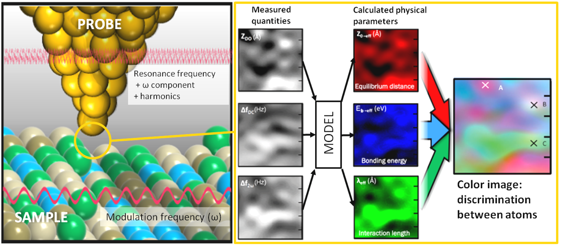 IMAGE: This is an examample of cilicon atoms represented in color.CREDIT:2017 HIDEKI KAWAKATSU, KAWAKATSU LABORATORY, INSTITUTE OF INDUSTRIAL SCIENCE< THE UNIVERSITY OF TOKYO.
MOVIE:This expains color AFM mechanism.